-
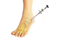
Ankle Arthroscopy
Ankle arthroscopy is a minimally invasive surgical procedure in which an arthroscope, a small, soft, flexible tube with a light and video camera at the end, is inserted into the ankle joint to evaluate and treat a variety of conditions.
-
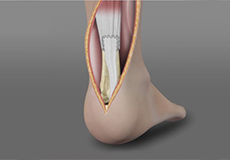
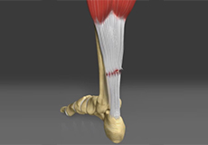
Achilles Tendon Repair
Tendons are the soft tissues connecting muscle to bone.The Achilles tendon is the longest tendon in the body and is present behind the ankle, joining the calf muscles with the heel bone.
-
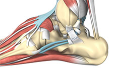
Ankle Ligament Reconstruction
Ankle ligament reconstruction may be performed arthroscopically under general anesthesia. Your surgeon will make small incisions in your ankle. A tiny camera and a few special instruments are inserted through the incisions to repair and strengthen the ligaments.
-
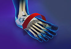
Ankle Instability Surgery
Ankle instability is a chronic condition characterized by the recurrent slipping of the outer side of the ankle. Instability is generally noticed during movement of the ankle joint, but can also occur while standing.
-
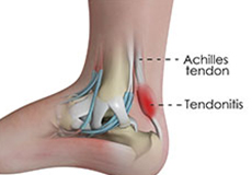
Achilles Tendinitis Treatment
Achilles tendinitis is the inflammation and irritation to the Achilles tendon, a strong fibrous cord present behind the ankle that connects your calf muscles to the heel bone.
-
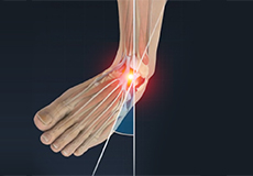
Ankle Sprain
A sprain is the stretching or tearing of ligaments. Ligaments connect adjacent bones and provide stability to a joint. An ankle sprain is a common injury that occurs when you suddenly fall or twist the ankle joint, or when you land your foot in an awkward position after a jump.
-
Ankle Instability
The joints of the ankle are held in place and stabilized by strong bands of tissue called ligaments. Ankle instability is a chronic condition characterized by a recurrent slipping of the outer side of the ankle.
-
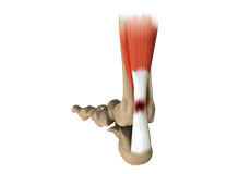
Achilles Tendon Rupture
The Achilles tendon is a strong fibrous cord present behind the ankle that connects the calf muscles to the heel bone. It is used when you walk, run and jump. The Achilles tendon ruptures most often in athletes participating in sports that involve running, pivoting and jumping.
-
Achilles Tendinitis
The Achilles tendon is a tough band of fibrous tissue that runs down the back of your lower leg and connects your calf muscle to your heel bone. The tendon is used when you walk, climb, jump, run and stand on your tip toes.
-
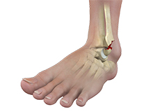
Ankle Fractures
Ankle injuries are very common in athletes and individuals performing physical work; often resulting in severe pain and impaired mobility. Pain after ankle injuries can either be from a torn ligament (ankle sprain) or broken bone (ankle fracture).
-
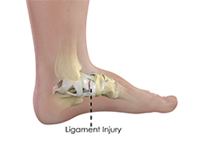
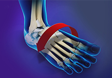
Ankle Ligament Injury
An ankle ligament injury, also known as an ankle sprain, can be caused by a sudden twisting movement of the foot during any athletic event or during daily activities. When stretched beyond its limit, the ligament may partially or completely tear.
What is the Normal Anatomy of the Foot and Ankle?
The foot and ankle form complex joints that are involved in movement and providing stability and balance to the body. The foot and ankle consist of 26 bones, 33 joints, and many muscles, tendons, and ligaments.
Bones of the Ankle
The ankle joint connects the leg with the foot and is composed of three bones: the tibia, fibula, and talus. The tibia or shinbone and fibula or calf bone are bones of the lower leg, which articulate with the talus or ankle bone, enabling up and down movement of the foot.
Three bony bumps present on the ends of the tibia and fibula form parts of the ankle joint:
- The medial malleolus, formed by the tibia, is found on the inside of the ankle.
- The posterior malleolus, also formed by the tibia, is found at the back of the ankle.
- The lateral malleolus, formed by the fibula, is found on the outer aspect of the ankle.
Bones of the Feet
The foot acts as a single functional unit, but can be divided into three parts: the hindfoot, midfoot and forefoot.
The hindfoot forms the ankle and heel, and is made up of the talus bone and calcaneus or heel bone. The heel bone is the largest bone in the foot.
The midfoot connects the hindfoot to the forefoot, and consists of one navicular bone, one cuboid bone, and three cuneiform bones. The navicular bone is found in front of the heel bone, and the cuneiform and cuboid bones are arranged in front of the navicular bone.
These bones are connected to five metatarsal bones of the forefoot that form the arch of the foot for shock absorption while walking or running. The forefoot is also made up of the toes or digits, formed by bones called phalanges - three in each toe, except the big toe, which has only two phalanges. The big toe has two additional tiny round sesamoid bones in the ball of the foot, which helps in upward and downward movements of the toe.
Ankle and Foot Joints
There are 33 joints in the ankle and foot. They include:
- Hinge joints in the ankle, which allow flexion (bending) and extension
- Gliding joints found in the hindfoot, which allow gliding movements
- Condyloid joints found in the forefoot and toes, which allow the flexion (bending) and extension, adduction, and abduction (sideward movement).
The joints of the foot and ankle provide stability and support the weight of your body, helping you to walk or run, and adapt to uneven grounds.
Soft Tissues of the Ankle and Foot
Our feet and ankle bones are held in place and supported by various soft tissues such as cartilage, ligaments, muscles, tendons, and bursae.
The joint surface of all the bones of the ankle and foot are lined by a thin, tough, flexible, and slippery surface called the articular cartilage, which acts as a shock absorber and cushion to reduce friction between the bones. The cartilage is lubricated by synovial fluid, which further enables smooth movement of the bones.
Ligaments are tough rope-like tissue that connect bones to other bones, and hold them in place, providing stability to the joints. The plantar fascia is the largest ligament in the foot, originating from the heel bone to the forefoot, it extends along the lower side of the foot and is involved in maintaining the arch of the foot. The plantar fascia ligament stretches and contracts to provide balance and strength to the foot. Lateral ligaments on the outside of the foot and medial ligaments on the inside of the foot provide stability and allow up and down movement of the foot.
The foot is made up of 20 muscles that help in movement. The main muscles include:
- Anterior tibial muscle, which allows up and down movement of the foot
- Posterior tibial muscle, which supports the arch
- Peroneal tibial muscle, which controls movement on the outside of the ankle
- Extensors, which enable the ankle to raise the toes just before stepping forward
- Flexors, which stabilize the toes against the floor
- Smaller muscles that help the toes to lift and curl
Tendons are soft tissues that connect muscles to bones. The largest and strongest tendon in the foot is the Achilles tendon, present at the back of the lower leg around the heel bone. Other tendons include peroneal and anterior and posterior tibialis.
Bursae are small fluid-filled sacs that decrease friction between tendons and bone or skin. They contain special cells called synovial cells that secrete a lubricating fluid.